Assalamualaikum.
Today we learn about PDB or Protein Data Bank. For your information, Protein Data Bank (PDB) is a repository for the three-dimensional structural data of large biological molecules, such as proteins and nucleic acids. The data, typically obtained by X-ray crystallography or NMR spectroscopy and submitted by biologist and biochemists from around the world, are freely accessible on the Internet via the websites of its member organisations (PDBe, PDBj,and RCSB). The PDB is overseen by an organization called the Worldwide Protein Data Bank , wwPDB. The PDB is a key resource in areas of structural biology , such as structural genomics. Most major scientific journals, and some funding agencies, such as the NIH in the USA, now require scientists to submit their structure data to the PDB. If the contents of the PDB are thought of as primary data, then there are hundreds of derived (i.e., secondary) databases that categorize the data differently. For example, both SCOP and CATH categorize structures according to type of structure and assumed evolutionary relations; GO categorize structures based on genes.
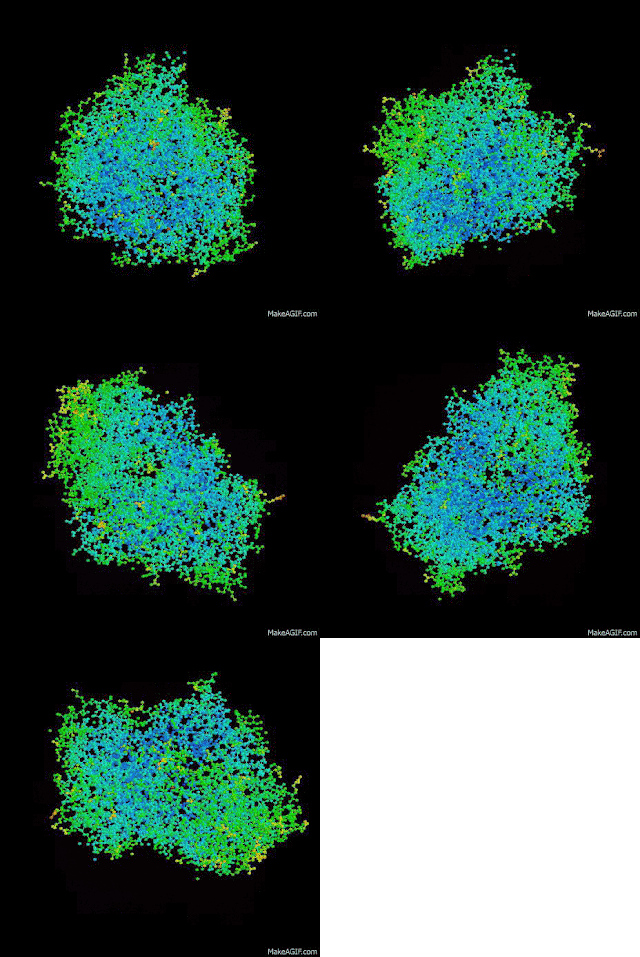
Here, the some example of protein structures:
1) Lipase
The binding of Thermomyces lanuginosa lipase and its mutants [TLL(S146A), TLL(W89L), TLL(W117F, W221H, W260H)] to the mixed micelles of cis-parinaric acid/sodium taurodeoxycholate at pH 5.0 led to the quenching of the intrinsic tryptophan fluorescence emission (300-380 nm) and to a simultaneous increase in the cis-parinaric acid fluorescence emission (380-500 nm). These findings were used to characterize the Thermomyces lanuginosa lipase/cis-parinaric acid interactions occurring in the presence of sodium taurodeoxycholate. The fluorescence resonance energy transfer and Stern-Volmer quenching constant values obtained were correlated with the accessibility of the tryptophan residues to the cis-parinaric acid and with the lid opening ability of Thermomyces lanuginosa lipase (and its mutants). TLL(S146A) was found to have the highest fluorescence resonance energy transfer. In addition, a TLL(S146A)/oleic acid complex was crystallised and its three-dimensional structure was solved. Surprisingly, two possible binding modes (sn-1 and antisn1) were found to exist between oleic acid and the catalytic cleft of the open conformation of TLL(S146A). Both binding modes involved an interaction with tryptophan 89 of the lipase lid, in agreement with fluorescence resonance energy transfer experiments. As a consequence, we concluded that TLL(S146A) mutant is not an appropriate substitute for the wild-type Thermomyces lanuginosa lipase for mimicking the interaction between the wild-type enzyme and lipids.
2) Neprilysin
Neutral endopeptidase (NEP) is the major enzyme involved in the metabolic inactivation of a number of bioactive peptides including the enkephalins, substance P, endothelin, bradykinin and atrial natriuretic factor. Owing to the physiological importance of NEP in the modulation of nociceptive and pressor responses, there is considerable interest in inhibitors of this enzyme as novel analgesics and antihypertensive agents. Here, the crystal structures of the soluble extracellular domain of human NEP (residues 52-749) complexed with various potent and competitive inhibitors are described. The structures unambiguously reveal the binding mode of the different zinc-chelating groups and the subsite specificity of the enzyme.
3)HTPX
4)LONA
Electrostatic interactions mediated by arginine residues in
AAA+ module of Brevibacillus theroruber Lon protease are critical for DNA
binding and allosteric stimulation
4)PROLY
The solution structure of the periplasmic cyclophilin type cis-trans peptidyl-prolyl isomerase from Escherichia coli (167 residues, MW > 18.200) has been determined using multidimensional heteronuclear NMR spectroscopy and distance geometry calculations. The structure determination is based on a total of 1720 NMR-derived restraints (1566 distance and 101 phi and 53 chi 1 torsion angle restraints). Twelve distance geometry structures were calculated, and the average root-mean-square (rms) deviation about the mean backbone coordinate positions is 0.84 +/- 0.18 A for the backbone atoms of residues 5-165 of the ensemble. The three-dimensional structure of E. coli cyclophilin consists of an eight-stranded antiparallel beta-sheet barrel capped by alpha-helices. The average coordinates of the backbone atoms of the core residues of E. coli cyclophilin have an rms deviation of 1.44 A, with conserved regions in the crystal structure of unligated human T cell cyclophilin [Ke, H. (1992) J. Mol. Biol. 228, 539-550]. Four regions proximal to the active site differ substantially and may determine protein substrate specificity, sensitivity to cyclosporin A, and the composite drug:protein surface required to inhibit calcineurin. A residue essential for isomerase activity in human T cell cyclophilin (His126) is replaced by Tyr122 in E. coli cyclophilin without affecting enzymatic activity.
5)LEXA
LexA
repressor undergoes a self-cleavage reaction. In vivo, this reaction
requires an activated form of RecA, but it occurs spontaneously in vitro
at high pH. Accordingly, LexA must both allow self-cleavage and yet
prevent this reaction in the absence of a stimulus. We have solved the
crystal structures of several mutant forms of LexA. Strikingly, two
distinct conformations are observed, one compatible with cleavage, and
the other in which the cleavage site is approximately 20 A from the
catalytic center. Our analysis provides insight into the structural and
energetic features that modulate the interconversion between these two
forms and hence the rate of the self-cleavage reaction. We suggest RecA
activates the self-cleavage of LexA and related proteins through
selective stabilization of the cleavable conformation.
There are many
websites that provide information about protein data bank. You can visit either
one of them
Lastly, below is the polymer and chain of the structures. Hope you all enjoy learning PDB. May Allah bless.
| Name | Polymer | Chain |
|---|---|---|
| LIPASE | 1 | A,B |
| NEPRILYSIN | 1 | A |
| HTPX | 1 | A,B |
| LONA | 1 | A,B |
| PROLYL | 1 | A |
| LEXA | 1 | A,B |


No comments:
Post a Comment